Total Pageviews

Jun 19, 2015

Classification of topical corticosteroids


 Topical corticosteroids have been ranked in terms of potency into four groups consisting of seven classes. Class I topical corticosteroids are the most potent and Class VII are the least potent. Efficacy and side-effects are greatest with the Class I ultra-high-potency preparations which should only be used for limited time periods (2-3 weeks)


----ULTRA HIGH POTENCY
      Class I
Clobetasol propionate Cream, 0.05%

------HIGH POTENCY

        1)  Class II
Amcinonide Ointment, 0.1%
Betamethasone dipropionate Ointment, 0.05%
 
Desoximetasone Cream or ointment, 0.025%
Fluocinonide Cream, ointment or gel, 0.05%
 
Halcinonide Cream, 0.1%

        2)  Class III
Betamethasone dipropionate Cream, 0.05%
Betamethasone valerate Ointment, 0.1%
 
Triamcinolone acetonide Ointment, 0.1%

-----  MODERATE POTENCY
        1) Class IV
Desoximetasone Cream, 0.05%
Fluocinolone acetonide Ointment, 0.025%
 
FludroxycortideOintment, 0.05%
Hydrocortisone valerate Ointment, 0.2%
 
Triamcinolone acetonide Cream, 0.1%

      2) Class V
Betamethasone dipropionate Lotion, 0.02%
Betamethasone valerate Cream, 0.1%
Fluocinolone acetonide Cream, 0.025%
 
FludroxycortideCream, 0.05%
Hydrocortisone butyrate Cream, 0.1%
 
Hydrocortisone valerate Cream, 0.2%
Triamcinolone acetonide Lotion, 0.1%

------------LOW POTENCY
        1) Class VI
Betamethasone valerate Lotion, 0.05%
Desonide Cream, 0.05%
 
Fluocinolone acetonide Solution, 0.01%

       2) Class VII
Dexamethasone sodium phosphate Cream, 0.1%
Hydrocortisone acetate Cream, 1%
 
Methylprednisolone acetate Cream, 0.25%

May 13, 2015

DRESS syndrome

DNB 2013

DRESS syndrome is associated with all except
a.Drug reaction
b.myocarditis
c.Eosinophilia
d.Encephalitis
Ans: d

DRESS syndrome- Short synposis

•Initially, 'anticonvulsant hypersensitivity syndrome' was first used to label this specific entity.

•Drug hypersensitivity syndrome (DHS) also referred to as DRESS (Drug Reaction with Eosinophilia and Systemic Symptoms) term is now used.

•It is an adverse drug reaction commonly associated with the aromatic antiepileptic drugs viz., phenytoin , carbamazepine , phenobarbital , lamotrigine, primidone etc. It may also be seen with Allopurinol, Dapsone, Minocycline, Nevirapine

•It is usually defined by the triad of fever, skin rash and symptomatic or asymptomatic internal organ involvement, within the first 2-8 weeks after initiation of therapy.

•Skin manifestation are in the form of exanthem, erythroderma or blisters. Internal manifestations include, among others, agranulocytosis, hepatitis, nephritis and myocarditis.

•Management mainly includes immediate withdrawal of the culprit drug, symptomatic treatment and systemic steroids or immunoglobulins.

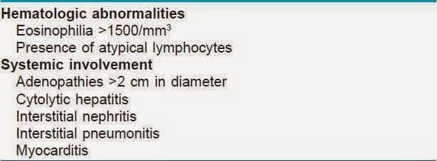
Features for DRESS are as below: 

Apr 8, 2015

Partner management of STDs (CDC Guidelines)


Partner management of STDs 
(CDC Guidelines)


    STD
Yes/No
                    Comments
Syphilis
Yes
- Partners to be treated if exposure within last 90 day before onset of pts symptoms (if early, secondary and early latent syphilis in the patient. VDRL > 1:32 indicates patient is in early syphilis stage)

- If exposure >90 days before onset of pts symptoms should be offered serology and if not possible, treat presumptively

Chancroid
Yes
Partners to be treated if exposure within last 10 day before onset of pts symptoms

Donovanosis
Yes
Partners to be treated if exposure within last 60 day before onset of pts symptoms

LGV
Yes
Partners to be treated if exposure within last 60 day before onset of pts symptoms

Chlamydia/
Gonorrhea (generally treated together)

Yes
Partners to be treated if exposure within last 60 day before onset of pts symptoms
Trichomonas
Yes
Partner treatment is always done (time before exposure to patient not specified)

PID
Yes
Partners to be treated if exposure within last 60 day before onset of pts symptoms

Epididymitis
Yes
Partners to be treated if exposure within last 60 day before onset of pts symptoms

Genital Scabies
Yes
Partners to be treated if exposure within last 30 day before onset of pts symptoms

Herpes genitalis

No
Partners evaluated for active disease, Asymptomatic partners NOT treated

Bacterial Vaginosis

No
NOT required
Candida
No
Treatment NOT done for asymptomatic partners as most infections are not sexually transmitted. Only for symptomatic male  partners with erythema, pruritus and irritation on glans


Mar 31, 2015

Acantholysis


Introduction

  • The term acantholysis  is derived from the Greek words akantha, meaning a thorn or prickle, and lysis, i.e. loosening.
  • Acantholysis is the term used to describe loss of cohesion between  keratinocytes, due to breakdown of intercellular bridges (eg- Desmosomes). This makes the normally polygonal keratinocytes  circular (Circular keratinocytes= Acantholytic cells). It results in the formation of intraepidermal clefts, vesicles and bullae.
Although acantholysis may occur at any level of the epidermis, the location of the blister is often used as a clue to the underlying disorder; for example, superficial (subcorneal) acantholysis favors pemphigus foliaceus, while acantholysis in the deeper aspects of the epidermis is more characteristic of pemphigus vulgaris

Acantholytic cell in pemphigus- Yellow arrow
(Ref: IJDVL. Seshadri et al.)

Primary Acantholysis

Acantholysis is either due to direct injury to desmosomes (eg: Pemphigus, bullous impetigo, staphylococcal scalded skin syndrome SSSS) or due to hereditary defects in their construction (eg: Darier's disease and Hailey-Hailey disease). Thus, in these diseases, acantholysis is the primary event leading to the formation of intra-epidermal cavities and hence the manifestations of the disease.  Eg: pemphigus, Darier’s disease etc 


Secondary Acantholysis

The acantholysis is secondary to alteration or damage to keratinocytes by various factors. In other words, keratinocytes are injured first followed by subsequent disintegration of desmosomes. Eg: In herpes, intraepidermal vesicle is secondary to ballooning degeneration of keratinocytes causing circular keratinocytes. Tzanck cells are multinucleated giant cells seen after fusion of individual acantholytic cells and is characteristically seen in herpes.


MCQs

1) A middle aged female presents with flaccid bullae in skin and oral erosions. Histopathology shows intra-epidermal acantholytic blisters. The most likely diagnosis is (PGI 05)
a) Pemphigus vulgaris
b) Bullous pemphigoid
c) Dermatitis herpetiformis
d) Epidermolysis bullosa
e) Pemphigus foliaceous
Ans: a

2) A female presents with persistent painful oral erosions with acantholytic cells. Most likely diagnosis (AI 08)
a) Dermatitis herpetiformis
b) Bullous pemphigoid
c) Pemphigus vulgaris
d) Epidermolysis bullosa
Ans: c

3) Acantholysis is seen in (PGI 99)
a) Bullous pemphigoid
b) Dermatitis herpetiformis
c) Hailey-Hailey disease
d) Darier’s disease
e) Pemphigus vulgaris
Ans: c.d.e

4) Acantholysis is seen in (AI 95)
a) Epidermis
b) Dermis
c) Dermoepidermal junction
d) Subcutaneous tissue
Ans: a

5) Acantholysis is due to destruction of (AIIMS 97)
a) Epidermis
b) Dermis
c) Basement membrane
d) Intercellular susbstance
Ans: d

6) Acantholytic cells are (SGPGI 01)
a) Epidermis cells
b) Plasma cells
c) Keratinocytes
d) Giant cells
Ans: c

7) Acantholytic cells are (PGI 96)
a) Cells with hyperchromatic nuclei and perinuclear halo
b) Cells with hypochromatic nuclei and perinuclear halo
c) Multinucleate cells
d) None
Ans: a

8) A 50 year old man with flaccid bullae and oral ulcers. Smear from skin lesions would show (AI 96)
a) Tzanck cells
b) Acantholytic cells
c) Necrosis
d) Koilocytes
Ans:b


Mar 17, 2015

Air Borne contact dermatitis (ABCD)


Air Borne contact dermatitis (ABCD)


Introduction
  • Parthenium hysterophorus and less commonly by other plants is one of the most intractable problems in dermatology in India
  • ABCD is a diagnosis that encompasses all photodermatoses predominantly of exposed parts of body, which are caused by substances released by plants which when released into the air, settle on the exposed skin and acted upon by UV light to cause disease.
  • In India, parthenium is also known as "Congress grass" or "Congress weed," which refers to the US congress (who allocated the shipment for Pune, India)
  • It is caused by airborne dry and friable plant particles including trichomes, and the most important allergens responsible for allergic contact dermatitis are sesquiterpene lactones (SQL)


Pathology

  • Combined type IV and type I hypersensitivity to parthenium has been postulated. 
  • It  is an immuno-inflammatory disease, which upon contact sensitization by parthenium antigen propagates as a cell-mediated hypersensitivity immune response with early sensitization phase and a subsequent elicitation phase, if antigen exposure persists.
 

Allergens in Parthenium dermatitis

  • The most important allergens responsible for allergic contact dermatitis is sesquiterpene lactones (SQL)
  • It is present in the leaf, stem, flower and pollen, but the highest concentrations of SQLs are present in the small glandular hairs (trichomes) present on the undersurface of the leaves and stem

Presentation

  • Most of the airborne contact dermatitis starts from the eyelids, suggesting that airborne allergens initially lodge there because of the skin folds and cause dermatitis, later affects the face, especially the eyelids and/or neck, V of the chest
  • A seasonal variation is initially observed with the dermatitis flaring in the summers corresponding to the growing season and disappearing in winters.
  • Repeated exposures over many years may result in widespread, extensive, and eventually chronic lichenified dermatitis that may persist throughout the year.
  • The involvement of both light-exposed and protected areas helps to differentiate ABCD from only sun induced eruptions ( photodermatitis
  Thickened lichenified skin (above)
Involvement of face, v of the neck

lichenified papules on dorsum of hand

Diagnosis

Patch testing with plant allergen is the simplest way of confirming parthenium contact allergy. 
    • Photopatch test positive (In photopatch test, parthenium area is irradiated with UV light)= If redness is seen it indicates Parthenium + Sun dermatitis (Phytophotodermatitis)
    look at redness on position no.2 on both sides

    Treatment


    Systemically, In India- Azathioprine is the commonest drug prescribed after initial steroid therapy, especially for chronic therapy in these usually long term diseases

    MCQ
    A 55 year old farmer with diabetes and hypertension gets 
    air borne contact dermatitis. The drug of choice is
    a) Corticosteroids
    b) Thalidomide
    c) Azathioprine
    d) Cyclosporine
    Ans: C