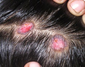
Erythematous scaly plaques with adherent scale, causing scarring alopecia. Notice the variable pigmentation with depigmentation in occipital area and hyperpigmented border (on right side)
In Brief
- Lupus erythematosus (LE) is usually divided into two main
types: DLE and SLE
- Discoid lupus erythematosus (DLE) generally occurs in young
adults, with women outnumbering men 2:1 (Discoid= Disc-like)
- DLE can be subdivided into a localized form (localized DLE) in which lesions are confined to the face above the
chin, the scalp and the ears, and a disseminated form (Disseminated
DLE) in which lesions go below the neck.
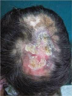
Presentation
- Discoid lesions are usually localized above the neck. Favored
sites are the scalp, bridge of the nose, malar areas, lower lip, and ears.
- Usually, lesions occur as well-defined erythematous plaques.
- There is adherent scale in many cases (as is seen in
photo) and when this is removed its undersurface shows horny plugs which have
occupied underlying dilated pilosebaceous canals (When not obscured by scaling,
these dilated follicular openings may be seen clinically as well). This
so-called ‘tin-tack’ sign or carpet tack sign or langue du
chat (cat’s tongue) sign.- MCQ
- Scarring alopecia ensues- (cause of scarring alopecia. SLE causes both scarring and Non-scarring alopecia) MCQ
- Itching and tenderness are common.
Investigation
1) Histology- Investigation of choice MCQ - 2 out of 3 should be
present
- Degenerative changes in the dermal connective tissue, consisting of
hyalinization, oedema and fibrinoid change in upper dermis. Thickening of the
basement membrane zone and dermal mucin deposition
- Liquefaction degeneration of the basal cell layer of the epidermis
- A patchy dermal lymphocytic infiltrate with a few plasma
cells and histiocytes, particularly around the appendages, which may be
atrophic
(Ref: http://pixshark.com/discoid-lupus-erythematosus-histopathology.htm)
(Ref: IJDVL)
2) ANA – Antinuclear antibodies are found
in between 5-60% of cases depending on patient selection and laboratory
techniques: the ‘homogeneous’ type of antinuclear factor being twice as
frequent as the ‘speckled’ type.
Treatment
Exposure to sunlight must be avoided, and a high
sun-protection factor (SPF) sunscreen should be used daily
Treatment chart for DLE
Topical
|
Systemic
|
|
Steroids
Potent or
superpotent topical corticosteroids are beneficial. The single most effective
local treatment is the injection of corticosteroids into the lesions.
Triamcinolone acetonide, 2.5–10 mg/mL, is infiltrated into the lesion through
a 30-gauge needle at intervals of 4–6 weeks.
|
1) Antimalarials
The safest class
2)
Acitretin
Second-line
agents and are particularly helpful in treating hypertrophic DLE
|
|
|
|
|
|
|
Complication
1) Rate of progression
to SLE- Extra edge point MCQ
- Localised DLE- 1.2% risk
- Disseminated DLE- 22% risk
- Females with DLE before< age of 40 years with HLA-B8, have
an increased risk
- Patients with DLE showing signs of nephropathy, arthralgia
and ANA titres of 1 : 320 or more should be carefully monitored
2) Rarely, aggressive squamous cell carcinoma arises in
long-standing lesions of DLE.
|


2 comments:
Sir what about its prognosis?
Rahul, DLE prognosis is favorable regarding mortality, however, morbidity can be considerable. It is a chronic disease with exacerbations and partial remissions. Scaring alopecia is permanent.
Post a Comment