Total Pageviews

Feb 27, 2015

How to decide level of Melanin in pigmentation disorders


Clinical Colour and level of melanin


See Table below
 
Level of colour
Clinical colour
Disease eg

a) Intraepidermal only
Black
Melanoma in situ
(lentigo maligna)
b) Basal epidermis/ Dermoepidermal junction
Brown
  • Lentigine
  • Junctional nevus
  • CafĂ©-au-lait macules
  • Melasma
c) Superficial dermis
Purple/Violaceous
  • Lichen planus
  • Fixed drug eruption
d) Deep dermis
Blue/Grey
  • Mongolian spot
  • Nevus of Ota
  • Nevus of ito
 
 Intraepidermal (Black)
Intraepidermal Lentigo melanoma (melanoma-in-situRef: dermnetz)


Basal epidermal and Dermo-epidermal Junction (Brown)

Junctional Nevus (Ref: Dermnetz)


Cafe-au-Lait macule in Neurofibromatosis


Lentigines on face


Melasma (Ref: DermQuest)


Superficial Dermal (purple/ violaceous)

Lichen Planus- Violaceus Papules (Ref: DermQuest)


Purple/ Violaceous papules in lichen planus


violaceous lesions in Fixed drug eruption (Ref: DermQuest)


Deep Dermal (blue/grey)


Nevus of Ota- This a congenital blue coloured nevus along ophthalmic and maxillary nerves (Ref: DermQuest)
Mongolian Spot This a congenital blue coloured nevus on lower back and sacrum (Ref:DermQuest)

Some tips in an MCQ
1) blue around eyes, on sclera and cheek since birth-unilateral is ALWAYS " nevus of ota"
2) blue on shoulder girdle since birth- ALWAYS "nevus of ito"
3) blue on lumbosacral region in a child since birth- ALWAYS "mongolian spot"
4) brown patch bilaterally on cheek- ALWAYS melasma/chloasma

Feb 24, 2015

Guttate psoriasis-photofeature


A child presents with few small erythematous papules with minimal scaling on trunk and arms. His parents report a recent sorethroat. What is the likely diagnosis?


Answer: Guttate Psoriasis

Expl: 

Introduction


  • It is derived from the Latin word gutta, meaning drop (Guttate psoriasis is a type of psoriasis in which there are small, separated teardrops like lesions)
  • The role of infection (tonsillar Streptococcus pyogenes infection) in precipitating guttate psoriasis is well known.  Infection often precedes the eruption by 2-3 weeks.


Clinical feature

  • It is particularly common in children
  • Common sites are the arms, legs, and torso.
  • Acute onset of small, 1-10 mm diameter, droplike, erythematous papules, usually with a fine scale (generally the classical silvery scale is not easily seen)


How does streptococcus cause Guttate psoriasis ?

  • Still under debate. 
  • It has been suggested that streptococcal M protein may be similar to keratin in skin and hence leads to a molecular mimicry immunological response towards keratinocytes instead of the original streptococcus. This cross- reactivity between streptococcal antigens and keratinocyte components could explain the initiation of a disease process. 


Treatment

  • There is no firm consensus on specific treatment algorithms
  • Antibiotics to eradicate streptococcus are often started. Eg: Ăź -lactum antibiotics, Erythromycin,rifampicin etc


Prognosis

  • Episodes may recur, especially those due to pharyngeal carriage of streptococci
  • Generally, the disease is self-limiting, but a certain percentage of cases progress to chronic plaque psoriasis.


Feb 19, 2015

Hypopigmented patch on face- how to decide diagnosis in an MCQ- Important MCQ


A year old girl presents with ill defined hypopigmented patches on cheek with fine scaling. Her mother has a history of asthma. 


The best possible diagnosis is
a) Indeterminate hansens
b) Pityriasis alba
c) Pityriasis versicolor
d) Atopic dermatitis

Ans: b

Expl:

         
Pointer words for diagnosis of hypopigmented patch on cheek in children


Look for these phrases-


Diagnosis
  • Single/ few hypopigmented patches on cheek with fine scaling.
  • No atrophy.
  • Normal sensations on patch
  • Personal/ family h/o atopy in the questions (eg: asthma, recurrent URTI, etc)
  • Lesions come and go
Pityriasis alba (pityriasis= powder)
  • Single/ Few hypopigmented patches on cheek.
  • No scaling
  • Lesions show mild atrophy
  • Sensations are usually normal
  • Child from endemic areas like Bihar/ UP/ West Bengal,Tamil Nadu, etc
Indeterminate hansens
  • Multiple hypopigmented macules/ patches with scaling on scratching (scratch sign). Usually MCQ will say “multiple lesions on chest and trunk”, sometimes face.
  • Perifollicular hypopigmentation (this word is often present in questions)
  • Can occur in multiple shades of colours (brown/ hypopigmentated/red/pink)
  • Woods lamp-greenish yellow fluorescence. KOH smear positive (spaghetti and meat ball appearance)
  • no h/o atopy in the question
Pityriasis versicolor
  • Depigmented patches with/without leucotrichia (white hair)
Vitiligo
  • Depigmented/hypopigmented circular macule over centre of forehead in a female
Leucoderma due to bindi (agent causing it is para-tertiary butyl phenol)


Why not P. versicolor in this MCQ- see pic below ?


Typical pityriasis versicolor
Note the number of lesions. On scratching there will be fine scaling
(Ref:ehow)


Typical pityriasis versicolor on face
Note the number of lesions. On scratching there will be fine scaling
(Ref:skinsight.com)


Why not Atopic dermatitis?

                                           Typical atopic dermatitis in children (Ref:www.salineherapy.com)

    Note erythema, oozing and inflammation
 (Atopic dermatitis is itchy, P.alba is not.There is no hypopigmentation in atopic dermatitis)

     
Also, for diagnosis of atopic dermatitis, these criteria have to be fulfilled: 


Typical Adult atopic dermatitis
Note the erythema, oozing and inflammation in flexure of knee
(Ref:www.derm-image.org)


Above: Typical change in sites of lesions in atopic dermatitis with age groups


Typical bindi leucoderma on central forehead


Note the depigmentation and white hairs


Feb 18, 2015

Discoid lupus erythematosus

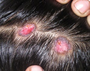
Erythematous scaly plaques on scalp, causing scarring alopecia. Notice the hyperpigmented borders and atrophic (=thin, hypopigmented) central area
Typical site- Auricular and Pre-auricular area
Erythematous scaly plaques with adherent scale, causing scarring alopecia. Notice the variable pigmentation with depigmentation in occipital area and hyperpigmented border (on right side)


In Brief


  • Lupus erythematosus (LE) is usually divided into two main types: DLE and SLE
  • Discoid lupus erythematosus (DLE) generally occurs in young adults, with women outnumbering men 2:1 (Discoid= Disc-like)
  • DLE can be subdivided into a localized form (localized DLE) in which lesions are confined to the face above the chin, the scalp and the ears, and a disseminated form (Disseminated DLE) in which lesions go below the neck.


Presentation


  • Discoid lesions are usually localized above the neck. Favored sites are the scalp, bridge of the nose, malar areas, lower lip, and ears.
  • Usually, lesions occur as well-defined erythematous plaques.
  • There is adherent scale in many cases (as is seen in photo) and when this is removed its undersurface shows horny plugs which have occupied underlying dilated pilosebaceous canals (When not obscured by scaling, these dilated follicular openings may be seen clinically as well). This so-called ‘tin-tack’ sign or carpet tack sign or langue du chat (cat’s tongue) sign.-    MCQ
  • Scarring alopecia ensues- (cause of scarring alopecia. SLE causes both scarring and Non-scarring alopecia) MCQ
  • Itching and tenderness are common. 


Investigation


1) Histology- Investigation of choice MCQ - 2 out of 3 should be present

  • Degenerative changes in the dermal connective tissue, consisting of hyalinization, oedema and fibrinoid change in upper dermis. Thickening of the basement membrane zone and dermal mucin deposition
  • Liquefaction degeneration of the basal cell layer of the epidermis
  • A patchy dermal lymphocytic infiltrate with a few plasma cells and histiocytes, particularly around the appendages, which may be atrophic


                                             (Ref: http://pixshark.com/discoid-lupus-erythematosus-histopathology.htm)


 (Ref: IJDVL)


2) ANA – Antinuclear antibodies are found in between 5-60% of cases depending on patient selection and laboratory techniques: the ‘homogeneous’ type of antinuclear factor being twice as frequent as the ‘speckled’ type. 


Treatment


Exposure to sunlight must be avoided, and a high sun-protection factor (SPF) sunscreen should be used daily


Treatment chart for DLE

Topical
Systemic

Steroids

Potent or superpotent topical corticosteroids are beneficial. The single most effective local treatment is the injection of corticosteroids into the lesions. Triamcinolone acetonide, 2.5–10 mg/mL, is infiltrated into the lesion through a 30-gauge needle at intervals of 4–6 weeks.
1) Antimalarials

The safest class

2) Acitretin

Second-line agents and are particularly helpful in treating hypertrophic DLE


Complication


1)  Rate of progression to SLE- Extra edge point MCQ


- Localised DLE- 1.2% risk
- Disseminated DLE- 22% risk
- Females with DLE before< age of 40 years with HLA-B8, have an increased risk
- Patients with DLE showing signs of nephropathy, arthralgia and ANA titres of 1 : 320 or more should be carefully monitored


2) Rarely, aggressive squamous cell carcinoma arises in long-standing lesions of DLE.